Quantifying functional effects of AFO alignment
Assessing device alignment is a key aspect of prosthetic practice, but when it comes to lower extremity orthotics the process is less common. That’s changing, however, as researchers begin to document the effects of alignment on gait biomechanics.
By Cary Groner, Lower Extremity Review July 2010
For decades, clinicians have speculated about the importance of alignment to the effectiveness of ankle-foot orthoses (AFOs). Rigorous approaches to measuring alignment and implementing protocols to optimize it have become generally available only relatively recently, however, and specialists continue to disagree about optimal methodology.
The type of condition being treated and the resources available to practitioners may lead to significant variations in practice as well. Those who specialize in post-stroke hemiplegia patients, for example, opt for different methods than those who most frequently treat children with cerebral palsy (CP). For that matter, it seems everyone could learn from the techniques of prosthetists, who have traditionally been more oriented toward dynamic alignment.
Regardless, the evolution and publication of increasingly detailed and flexible algorithms hold the promise of treatment plans that increasingly take into account the patient’s condition and individual needs.

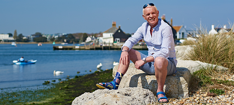
The SAFO (silicone ankle foot orthosis) is a skin-friendly, custom-fabricated silicone orthosis. As a supplement to orthotic fittings, users with foot drop can wear the SAFO without a shoe at home, in wet areas and even when swimming. Otto Bock
| Considerations for stroke patients |
Paul Charlton, an orthotist with Peacocks Medical Group in Newcastle-upon-Tyne, England, and chairman of the U.K. chapter of the International Society of Prosthetics and Orthotics (ISPO), has a special interest in adult neurology, particularly the rehabilitation of stroke patients.
“If the patient has the potential to improve their motor deficits, you want to give them an opportunity to get back to normality,” he said. “Physical therapists often destabilize them with their hands, to help them recruit the muscles they need to progress. I think there is a huge potential to use AFOs to achieve that, as an adjunct to therapy. Then, as patients progress and gain the ability to recruit their quadriceps and glutes, the orthosis can become more of a walking aid to help maintain that alignment.”
In most cases, Charlton tries to adjust alignments so that the tibia is pushed forward, bringing the knee out of hyperextension. This recruits antigravity muscles such as the quadriceps, glutes, and hip extensors, and supports more normal moments in the knee and hip.
Rigid AFOs are usually more appropriate for these patients than hinged ones, in Charton’s view.
“If a patient is uncomfortable stretching the calf, they may flex the knee instead, and to do that in stance they have to dorsiflex the ankle a little. A rigid AFO stops that dorsiflexion and helps keep the knee extended, which provides a better stretch of the gastrocnemius,” he said.
The authors of a 2001 paper in Topics in Stroke Rehabilitation took a comprehensive look at the orthotic management of patients with post-stroke hemiplegia.[1] Their considerations for orthotic management included seven domains: range of motion; gait assessment data; sensation / proprioception status; magnitude of spasticity; previous management; medical team feedback; and patient expectations. They also noted that orthotic goals included stance phase stability, foot clearance, foot prepositioning, adequate step length, and energy conservation.
“After the 1970s, when we went from metal to polymer AFOs, we lost some of the emphasis on looking at the alignment of the AFO as it fit in the shoe, and what that does to the person’s gait pattern,” said Bryan Malas CO MHPE, a coauthor of that study and now director of orthotics/prosthetics at Children’s Memorial Hospital in Chicago. “It’s important to see how the alignment sets the person up for the initial part of the gait cycle, then use that alignment to facilitate a better step length. I think that if you incline the tibia anteriorly several degrees, you put both the femur and trunk in better alignment.”Malas noted that when stroke patients walk without an orthosis, they often are plantar flexed at the ankle joint, which results in a toe-heel gait pattern and hyperextension of the knee. They then try to compensate for lost momentum with a forward trunk lean.
“In some of those patients, a small lift inside the shoe, under the AFO, gives them a much more upright posture,” Malas explained. “It also seems to facilitate better step length.”
In the article, Malas and his coauthor, Marty Kacen CPO, noted that when an orthosis is in the shoe, it should be checked in its relationship to the floor; once this is established, desired alignment can be implemented in dorsiflexion, neutral (tibia at 90° to the floor), and/or plantarflexion.
A slightly dorsiflexed alignment, for example, might be indicated for swing phase clearance or to increase a knee flexion moment during stance phase. Reasons for orthosis plantar flexion include facilitation of a knee extension moment during stance phase in the presence of knee instability.
“Clinically, I rely a great deal on feedback from the individual,” Malas added. “You first have to determine the ankle angle alignment of the orthosis based on the patient’s existing range of motion. Once that’s established and maintained, you go to the outside of the orthosis—I’m thinking of a solid AFO now—to the shoe, to garner functional gains and improve stability.”
“Articulated AFOs are probably all right if patients have the existing range of motion,” he said. “But if the patient does not have the necessary ankle range of motion and the AFO is allowed to articulate, there is the risk that patient may exhibit midfoot pronation to compensate for the lack of ankle range of motion. In such cases the heel-sole differential should be considered as well as the possibility of a solid AFO.”
| New angles on old problems |
A 2009 study published in Archives of Physical Medicine and Rehabilitation, however, found that changing AFO alignment did not significantly affect knee moments in patients with post-stroke hemiplegia.[2] The lead author of that paper was Stefania Fatone PhD BPO(Hons), a research assistant professor in the department of physical medicine and rehabilitation at Northwestern University’s Feinberg School of Medicine (Malas was one of her coauthors).
Fatone and colleagues compared articulated AFOs in two different alignment configurations. One condition involved a conventionally aligned AFO, with a plantar flexion stop set at a tibia-foot angle of 90°; when placed in a shoe, the shank was inclined anteriorly 5° to 7°. For the second condition, the plantar flexion stop was adjusted so that the shank was vertical in the shoe, accounting for the effect of the shoe heel height.
Both conditions significantly altered knee moment during the first half of stance from an external extensor moment to a flexor moment compared to a shoe-only condition, particularly in patients who walked with knee hyperextension while brace-free. However, the two alignment configurations did not differ significantly from each other, nor did either one eliminate hyperextension completely.
The plantar flexion stop creates an external flexor moment by blocking ankle plantar flexion. That way, Fatone told LER, the heel rather than the forefoot hits the ground first, and the ground reaction force is oriented less anterior to the knee joint.
“We showed it was possible to align the AFO to alter the moment acting at the knee for people with hyperextension as a result of their stroke,” she said. “The fact that it was a plantar flexion stop, that didn’t allow the tibia to recline beyond the position the AFO kept it at, was what influenced the moment.”The researchers had hypothesized that the initial internal flexor moment would be lower in the more vertically aligned orthosis; they concluded that the 5° to 7° change in alignment may not have been large enough to have a measurable effect on knee moments in the group of subjects studied.
“Those two alignments didn’t seem to have a drastically different effect on the moment profile in articulated AFOs,” Fatone said. “But the two positions we used weren’t necessarily sufficient changes in alignment to completely ameliorate the hyperextension that was happening, and I think one reason for that is maybe the tibia wasn’t sufficiently pitched forward in the shoe. Additionally, our articulated AFOs did not control forward progression of the tibia in mid to late stance, which may also affect knee moments.”
Fatone noted that for years the rule of thumb was that the patient’s ankle angle should be 90°; that is, with the foot parallel to the floor and perpendicular to the tibia, or shank. The work of researchers such as Elaine Owen, in England, has begun to change that idea with regard to rigid AFOs.
“It’s more complicated, and 90° may not be the right point if you are trying to influence things like moments at the knee,” Fatone continued. “You choose your ankle angle based on available muscle length, then once you’ve decided what’s optimal for that individual, you choose where you want the shank to be. You adjust the shank by using wedges under the AFO or even under the shoe, so you can separate the ankle angle from the shank angle, and you determine the latter with dynamic evaluation of the patient.”
A patient could have a tibia inclined anterior to vertical, in other words, while maintaining a 90° relationship between the tibia and the foot. Owen’s work in children with CP and spina bifida suggested an optimal (though individually variable) shank-to-vertical angle of roughly 10° anterior.
| Dynamic evaluations |
The kind of dynamic evaluation Fatone mentioned is more common in prosthetic fitting than in orthotics, but the evolution of the field may bring them closer together.
“Orthotists are now having to do some of the same dynamic alignments that prosthetists have always done,” she said. “That’s not a stretch if you were taught P and O as a double discipline, but orthotists who’ve never been exposed to dynamic alignment need to learn how to do it.”
When prosthetists fit an artificial leg, for example, they first put it together on the bench. Then, when they fit it to the patient, they make adjustments as the person is standing, then further tune it during walking. The goal is to manipulate several variables to produce a smooth, comfortable gait.
“If a person is wearing a rigid AFO, you can even tune their walk by changing the rockers on the shoe,” Fatone said. “But how do you do that? The rocker replaces some of what the ankle was doing, and its shape can determine a number of different things. The technology is making all this a more complex part of what we have to do, as orthotists.”
| Addressing issues in children with CP |
Fatone is originally from Melbourne, Australia, which increasingly seems a hotbed of orthotic innovation. A young researcher from the same city, Emily Ridgewell BPO(Hons), a doctoral candidate at the National Center for Prosthetics and Orthotics at Latrobe University, presented a paper at the May ISPO conference in Germany investigating the effect of alignment changes on solid and hinged AFOs in children with CP.[3] She and her coauthors reported that children studied had improved external knee extension moments and other beneficial effects as a result of increasing the shank-to-vertical angle of an AFO and footwear.
“We did three-dimensional gait analysis and looked at vertical shank alignment at five, ten, and fifteen-degree anterior tilts,” Ridgewell said, noting that the paper described a small pilot study that will soon be ramped up to a larger one with two cohorts of 20 patients each. Patients in one group will wear solid AFOs; those in the other group will have hinged AFOs with a plantar flexion stop. Researchers will adjust the heel-sole differential with wedges to incline the shank and move the knee joint closer to the ground reaction force vector, hence affecting knee moments.
“We can improve knee kinetics by either shifting the body toward the ground reaction force or by shifting the force posteriorly,” Ridgewell explained. “I don’t liken this to the complicated work of Elaine Owen, but there are lots of facilities that don’t have the resources, the gait labs, the people, or the time to run the tuning process like she does. I hope that our findings can be a resource to improve the gait of kids with CP with AFOs in places where they don’t have an entire tuning process.”
When clinicians discuss issues related to AFO alignment and effectiveness, Elaine Owen’s name and research comes up repeatedly.[4] She’s a pioneer in the field, having developed methodologies and algorithms that have begun to codify approaches to what is, for some, a complex and baffling task. Owen, who holds a Master’s of Science degree, is the superintendent pediatric physical therapist at the Child Development Center in Bangor, North Wales, U.K. In her research, she uses a video vector gait lab, which, she says, allows her to do a lot of work much quicker than she could with a 3-D lab.

Gait Analysis Lab, Shriners Hospital in Sacramento
“I teach people about normal gait,” she said. “How segments move, how joints move, how kinetics are created. Looking at those three components and how the distal movements affects the proximal movements is the key. Children with disabilities have distal problems—weak or stiff muscles—that create distal gait abnormalities, and because they have abnormal neurology, they produce all kinds of abnormal compensations. The point is to start them walking normally from a very young age, and the trick to that is to understand normal gait very well, then replicate it.”
Owen uses the term “segment” to mean each of the individual sections—foot, shank, thigh—that are connected by joints. The alignment of a segment is the spatial relationship between the ends of the segment and the alignment of a given joint. She believes that clinicians must first normalize the kinematics of the shank segment, that this doesn’t require a normally moving ankle, and that achieving this usually requires a rigid AFO combined with appropriate footwear designs.
“People have been aligning orthoses from traditional beliefs of how to align them rather than based on good research,” she said. “These children need more inclined alignment of the AFO-footwear combination than is usually provided, and footwear design is an important part of the prescription.”
Owen has produced graphic algorithms for making design and alignment decisions that will be published in Prosthetics and Orthotics International this September.[5]
“In cerebral palsy patients, you have to get the ankle angle right to reduce contractures,” she explained. “You have to stretch the muscle not too little, not too much when they’re walking. You want the knees to straighten in gait; you don’t want the foot to pronate or supinate in the AFO; you have to account for the bony structure of the foot, because if you get a wrong pull on the gastroc it will deform the foot bones.”
Owen’s first algorithm, then, relates to the ankle angle, between the shank and the foot. The second is for designing, aligning, and tuning the AFO-footwear combination. Tuning, as defined by Owen, is “adjusting to optimize.”
“You’ve got to fully tune the whole combination with all the variables in three planes,” she said. “You have to address heel design, pitch, and sole design. It’s very important to get ground reaction force vectors and kinetics right; if you don’t, it can create contracture and deformity as the child grows.”
Owen likens all the possible design options for the AFO and the footwear to a jigsaw; for each child, the clinician is trying to pick the right pieces and put it together.
“The algorithm is all about how you pick which bits for which children, based on walking patterns,” she said. “We’ve got to work quickly and accurately, because they only have one childhood.”
| Cary Groner is a freelance writer based in the San Francisco Bay area. |
Source Lower Extremity Review
| References |
- Orthotic management in patients with stroke, Malas B, Kacen M. Top Stroke Rehabil. 2001 Winter;7(4):38-45.
- Effect of ankle-foot orthosis alignment and foot-plate length on the gait of adults with poststroke hemiplegia, Fatone S, Gard SA, Malas BS. Arch Phys Med Rehabil. 2009 May;90(5):810-8. doi: 10.1016/j.apmr.2008.11.012.
- The biomechanical and functional effects of ankle foot orthosis shank-to-vertical angle in children with cerebral palsy: a pilot study, Ridgewell E, Gibson S, Nguyen T, Walker L. Presented at 13th ISPO World Conference, Leipzig, Germany, May 12–15, 2010.
- Shank angle to floor measures of tuned ankle-foot orthosis footwear combinations used with children with cerebral palsy, spina bifida and other conditions, Owen E. Gait & Posture 2002;16(Suppl 1):S132–133.
- The importance of being earnest about shank and thigh kinematics especially when using ankle-foot orthoses, Owen E. Prosthet Orthot Int. 2010 Sep;34(3):254-69. doi: 10.3109/03093646.2010.485597.

