
Dynamic bracing of pectus carinatum: a quantitative analysis
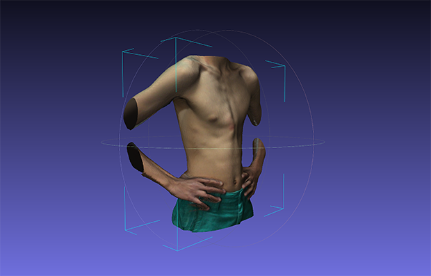
3D Scan, Braceworks® 2016.
See you at the 49th Annual Meeting of the Canadian Association of Paediatric Surgeons, CAPS in Banff, Alberta, October 5-7, 2017 at the Fairmont Banff Springs.
| Purpose |
Primary treatment of pectus carinatum (PC) is performed with an external brace that compresses the protrusion. Patients are ‘prescribed’ a brace tightening force. However, no visual guides exist to display this force magnitude. The purpose of this study was to determine the repeatability of patients in applying their prescribed force over time and to determine whether the protrusion stiffness influences the patient-applied forces and the protrusion correction rate.
| Methods |
Twenty-one male participants (12-17years) with chondrogladiolar PC were recruited at the time of brace fitting. Participants were evaluated on three visits: fitting, one month postfitting, and two months postfitting. Differences between prescribed force and patient-applied force were evaluated. Relationships of patient-applied force and correction rate with protrusion stiffness were assessed.
| Results |
Majority of individuals followed for two months (75%) had a significantly different patient-applied force (p<0.05) from their prescribed force. Protrusion stiffness had a positive relationship with patient-applied force, but no relationship with correction rate.
| Conclusion |
Patients did not follow their prescribed force. Magnitudes of these differences require further investigation to determine clinical significance. Patient-applied forces were influenced by protrusion stiffness, but correction rate was not. Other factors may influence these variables, such as patient compliance.
| References |
Bracing of pectus carinatum: A quantitative analysis, Bugajski T, Murari K, Lopushinsky S, Schneider M, Ronsky J. J Pediatr Surg. 2018 May;53(5):1014-1019. doi: 10.1016/j.jpedsurg.2018.02.034. Epub 2018 Feb 13.
Bracing of Pectus Carinatum: A Quantitative Analysis, Tomasz Bugajski. Thesis, Graduate Program in Biomedical Engineering, University Of Calgary, Alberta 2017
Dynamic bracing of pectus carinatum: a quantitative analysis, Bugajski T, Murari K, Lopushinsky S, Schneider M, Reichbart J, Ronsky J. Abstract 19, Canadian Association Of Paediatric Surgeons CAPS 49th Annual Meeting Banff, Alberta Canada, October 5-7, 2017
| Further reading |
The Dynamic Compression Brace for Pectus Carinatum: Intermediate Results in 286 Patients, de Beer SA, Gritter M, de Jong JR, van Heurn ELW. Ann Thorac Surg. 2017 Jun;103(6):1742-1749. doi: 10.1016/j.athoracsur.2016.12.019. Epub 2017 Mar 6.
A less intensive bracing protocol for pectus carinatum, Wahba G, Nasr A, Bettolli M. J Pediatr Surg. 2017 Nov;52(11):1795-1799. doi: 10.1016/j.jpedsurg.2017.01.057. Epub 2017 Jan 31.
A Simplified Method for Three-Dimensional Optical Imaging and Measurement of Patients with Chest Wall Deformities, Szafer D, Taylor JS, Pei A, de Ruijter V, Hosseini H, Chao S, Wall J. J Laparoendosc Adv Surg Tech A. 2019 Feb;29(2):267-271. doi: 10.1089/lap.2018.0191. Epub 2018 Sep 12.
Reconstruction of laser-scanned 3D torso topography and stereoradiographical spine and rib-cage geometry in scoliosis, Poncet P, Delorme S, Ronsky JL, Dansereau J, Clynch G, Harder J, Dewar RD, Labelle H, Gu PH, Zernicke RF. Comput Methods Biomech Biomed Engin. 2000;4(1):59-75.
Clinical impact of optical imaging with 3-D reconstruction of torso topography in common anterior chest wall anomalies, Poncet P, Kravarusic D, Richart T, Evison R, Ronsky JL, Alassiri A, Sigalet D. J Pediatr Surg. 2007 May;42(5):898-903. doi: 10.1016/j.jpedsurg.2006.12.070.