
Evaluation of the treatment of pectus carinatum with compressive orthotic bracing using three dimensional body scans
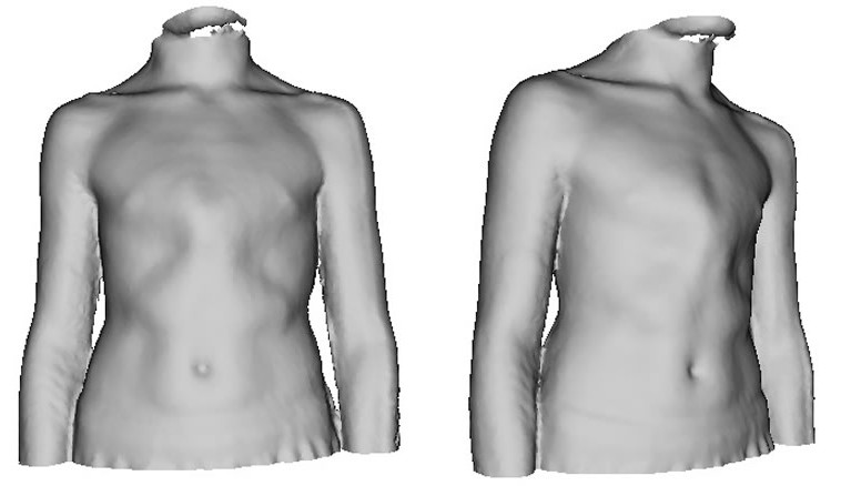
| 3D model used to assess and measure severity of pectus deformity demonstrating bilateral rib flattening in a severe symmetric pectus carinatum deformity. Ian Hunt, Pectus Clinic, Spire St. Anthony’s Hospital, London UK |
| Purpose. |
The purpose of this study is to measure the effectiveness of compressive orthotic brace therapy for the treatment of pectus carinatum using an adjusted Haller Index (HI) measurement calculated from 3D body scan (BS) images.
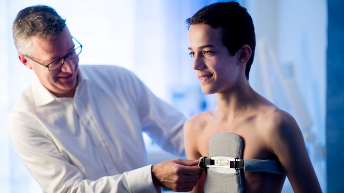
The low-profile light weight brace used by the Pectus Clinic is custom built on-site following extensive measurements and then re-checked following bracing to guarantee an ideal fit. At further follow-ups, the brace will be altered as the pectus carinatum remodels to give the best long-term results. Spire St. Anthony’s Hospital, North Cheam, Surrey. Photo credit PA
| Methods |
Pediatric patients with pectus carinatum were treated with either compressive orthotic bracing or observation. An adjusted BS Haller index (HI) was calculated from serial 3D BS images obtained on all patients. Medical records were evaluated to determine treatment with bracing and brace compliance more than 12 hours daily. Compliant patient measurements were compared to non-compliant and non-brace groups.
| Results |
Forty patients underwent compressive orthotic bracing, while ten were observed. Twenty-three patients were compliant with bracing, and seventeen patients were non-compliant. Compliant patients exhibited an 8.2% increase, non-compliant patients had a 1.5% increase, and non-brace patients exhibited a 2.5% increase in BS HI. The change in BS HI of compliant patients was significantly different compared to non-brace patients (p = 0.004) and non-compliant patients (p < 0.001).
| Conclusions |
Three dimensional BS is an effective, radiation free, and objective means to evaluate patients treated with compressive orthotic bracing.
| References |
Evaluation of the treatment of pectus carinatum with compressive orthotic bracing using three dimensional body scans, Wong KE, Gorton GE 3rd, Tashjian DB, Tirabassi MV, Moriarty KP. J Pediatr Surg. 2014 Jun;49(6):924-7. doi: 10.1016/j.jpedsurg.2014.01.024. Epub 2014 Feb 3.
| Further reading |
External Compressive Bracing With Initial Reduction of Pectus Carinatum: Compliance Is the Key, Fraser S, Harling L, Patel A, Richards T, Hunt I. Ann Thorac Surg. 2020 Feb;109(2):413-419. doi: 10.1016/j.athoracsur.2019.08.026. Epub 2019 Sep 23. Full text
Effectiveness of Compressive External Bracing in Patients with Flexible Pectus Carinatum Deformity: A Review, Hunt I, Patel AJ. Thorac Cardiovasc Surg. 2020 Jan;68(1):72-79. doi: 10.1055/s-0039-1687824. Epub 2019 Apr 25.
Initial reduction of flexible pectus carinatum with outpatient manipulation as an adjunct to external compressive bracing: technique and early outcomes at 12 weeks, Fraser S, Richards T, Harling L, Patel AJ, Hunt I. J Pediatr Surg. 2020 Jul;55(7):1347-1350. doi: 10.1016/j.jpedsurg.2019.09.024. Epub 2019 Nov 1.
Application of Reverse Engineering in Supporting the Treatment of Pectus Carinatum, Magdalena Antonowicz, Anita Kajzer, Wojciech Kajzer. Chapter, Information Technologies in Medicine, Volume 472 of the series Advances in Intelligent Systems and Computing pp 217-225. Springer
Congenital Thoracic Wall Deformities – Diagnosis, Therapy and Current Developments, Editor: Anton H. Schwabegger MS MSc Assoc Prof. Springer. ISBN: 978-3-211-99137-4 (Print) 978-3-211-99138-1 (Online)