
Does overgrowth of costal cartilage cause pectus carinatum?
A three-dimensional computed tomography evaluation of rib length and costal cartilage length in patients with asymmetric pectus carinatum.
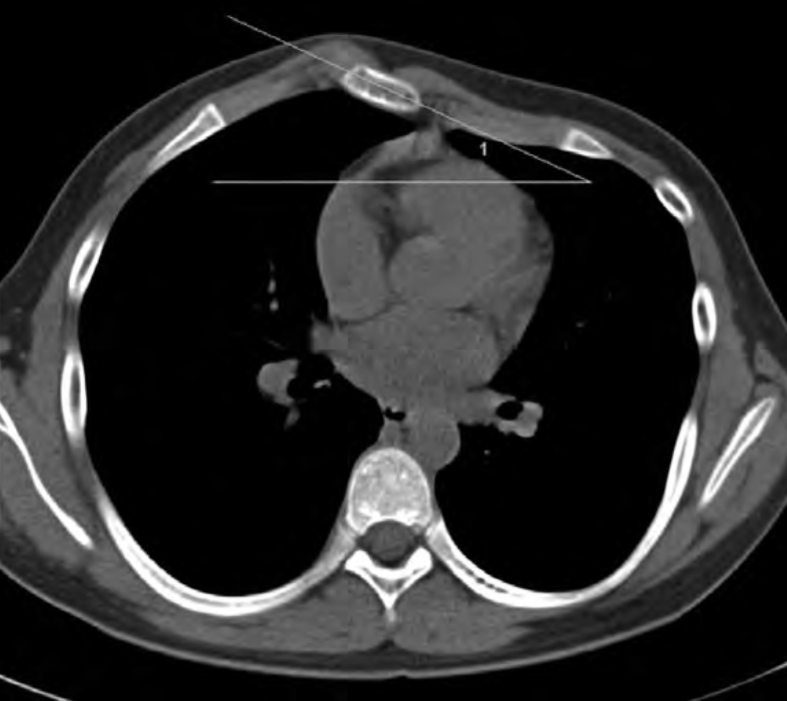
Figure 1. Measurement of the sternal rotation angle. The sternal rotation angle was measured as the maximum angle of the sternal slope against the baseline of the thorax. Patients with a >10° angle of sternal rotation were regarded as having asymmetric pectus carinatum. In this patient, the sternal rotation angle is 27° and the right side is the more protruded than the left. C.H. Park et al.
| OBJECTIVES |
| To evaluate whether the overgrowth of costal cartilage may cause pectus carinatum using three-dimensional (3D) computed tomography (CT). |
| METHODS |
| Twenty-two patients with asymmetric pectus carinatum were included. The fourth, fifth and sixth ribs and costal cartilages were semi-automatically traced, and their full lengths were measured on three-dimensional CT images using curved multi-planar reformatted (MPR) techniques. The rib length and costal cartilage length, the total combined length of the rib and costal cartilage and the ratio of the cartilage and rib lengths (C/R ratio) in each patient were compared between the protruding side and the opposite side at the levels of the fourth, fifth and sixth ribs. |
| RESULTS |
| The length of the costal cartilage was not different between the more protruded side and the contralateral side (55.8 ± 9.8 mm vs 55.9±9.3mm at the fourth, 70±10.8mm vs 71.6±10.8mm at the fifth and 97.8±13.2mm vs 99.8±15.5mm at the sixth; P>0.05). There were also no significant differences between the lengths of ribs. (265.8 ± 34.9 mm vs 266.3 ± 32.9 mm at the fourth, 279.7 ± 32.7 mm vs 280.6 ± 32.4 mm at the fifth and 283.8 ± 33.9 mm vs 283.9 ± 32.3 mm at the sixth; P > 0.05). There was no statistically significant difference in either the total length of rib and costal cartilage or the C/R ratio according to side of the chest (P > 0.05). |
| CONCLUSIONS |
| In patients with asymmetric pectus carinatum, the lengths of the fourth, fifth and sixth costal cartilage on the more protruded side were not different from those on the contralateral side. These findings suggest that overgrowth of costal cartilage cannot explain the asymmetric protrusion of anterior chest wall and may not be the main cause of pectus carinatum. |
Does overgrowth of costal cartilage cause pectus carinatum? A three-dimensional computed tomography evaluation of rib length and costal cartilage length in patients with asymmetric pectus carinatum, Park CH, Kim TH, Haam SJ, Lee S. Interact Cardiovasc Thorac Surg. 2013 Nov;17(5):757-63. doi: 10.1093/icvts/ivt321. Epub 2013 Jul 17.
Also see
Pectus Carinatum: To Brace or Not to Brace—A Picture Is Worth 1770 Words, Deepika Nehra MD, Sigmund H. Ein MD, Mark Tlumacki CO, Peter T. Masiakos MD. JPO 2009 Vol. 21, Num. 3 pp. 167-170