
Embedded 3D videos enhance visualization in Journal of X-Ray Science and Technology
The current issue of the Journal of X-Ray Science and Technology now features 3D imaging embedded within its articles for the first time. Use of this digital and multimedia technique enhances the value of five papers, which focus on kidney injuries, tumor diagnoses, and dental medicine. Videos of these rotating 3D images can be accessed directly, allowing readers to examine them in detail and gain deeper understanding of the diagnoses, prognoses, and treatments described.
| Diagnosis Value of Multi-Slice Spiral CT in Renal Trauma. Contrast-enhanced CT can quickly determine the condition of the renal parenchymal damage both in the arterial phase and venous phase, and clearly shows the range and the degree of renal injury and the renal pedicle vessels. The enhanced CT provides an accurate visualization of the kidney injury, which is transformed into 3D images, providing more accurate and intuitive images. Journal of X-Ray Science and Technology |
IOS Press November 21, 2016
Guest Editor Yuanyuan Zhang MD PhD of the Wake Forest Institute for Regenerative Medicine, Wake Forest University School of Medicine, Winston-Salem, North Carolina, USA, notes that “State-of-the-art cross-sectional imaging approaches make it possible to visualize diseases-affected tissues or defected organs with greater assurance by minimizing the interrupter of overlying tissues to focus on individual organs, which aids in the detection and characterization of targeted tissues. The rotated multimedia 3D videos provide animations, which could better help medical students, residents, practitioners, and inquisitive minds better understand the diseases.”
Five papers in this issue demonstrate how imaging techniques contributed to positive patient outcomes. The 3D imaging incorporated directly into the articles provides greater insights and learning opportunities.
Trauma to the kidney is common. While ultrasound and MRI can be used to evaluate these injuries, computed tomography (CT) is more effective, and 3D reconstructed CT is even more helpful. Physicians from the New District Central Hospital, Shenzhen, China, and the Wake Forest University School of Medicine, Winston-Salem, North Carolina, USA, examined 126 patients with kidney injuries, using multi-slice spiral CT (MSCT) between January 2012 and February 2016. MSCT achieved a 100% diagnostic accuracy rate, confirmed by surgical findings. The authors concluded that the enhanced MSCT scan permits reliable detection of renal trauma and the associated organ or tissue injuries, providing important clinical value for the diagnosis and classification of renal trauma or internal organ injures.
Wilm’s tumor or nephroblastoma, a rare kidney cancer commonly affecting children under age 5, can grow significantly without causing pain or other symptoms. Conservative renal surgery is the most commonly selected treatment, requiring accurate mapping of the tumor. This article describes how creation of a 3D image helped physicians remove a large tumor in a 3-year old girl, who is now in good health eight months after surgery.
A rare congenital disorder that results in an “extra” ureter in the genitourinary tract can be visualized more effectively using MSCT. In the next contribution, researchers describe the case of a 27-year-old male patient with an almost complete duplication of the ureter from his left kidney. Finding this structure and correcting it surgically was facilitated by a 3D reconstruction of the patient’s anatomy before surgery.
Adrenocortical carcinoma (ACC) is an extremely rare disease caused by a cancerous growth in the adrenal cortex. ACC is malignant and necessitates surgical removal. In the next article, physicians describe how they achieved a better surgical outcome by integrating 3D-reconstructed CT images into a dynamic video for preoperative planning and intraoperative guidance. They were able to resect the diseased adrenal gland completely without neighbor organ injury and surgical complications.
The final article concerns a not-so-rare condition, malocclusion or “poor bite.” Because many cases of malocclusion are found in young children, for whom the higher radiation dose and extended scanning time of standard CT scanning are not desirable, the authors describe a lower dose and faster technique, cone beam computed tomography (CBCT). With the advent of CBCT, it is now possible for clinicians to evaluate the hard and soft tissues of the maxillofacial region in 3D and high spatial detail.
According to Advisory Editor-in-Chief Hong Liu PhD, Center for Bioengineering and School of Electrical and Computer Engineering, University of Oklahoma, “Digital and multimedia techniques are used more and more to facilitate scientific presentations. As an interdisciplinary journal publishing numerous papers spanning the field of medical imaging, the Journal of X-Ray Science and Technology should lead the way to serve our authors and readers with advanced presentation methods.”
![]() Source IOS Press via EurekAlert! AAAS
Source IOS Press via EurekAlert! AAAS
| Featured articles |
Multimedia presentation assisted clinical diagnosis, prognosis and treatment, Yuanyuan Zhang MD PhD, Guest Editor and Hong Liu PhD, Editor-in-Chief. Journal of X-Ray Science and Technology, vol. 24, no. 5, pp. 647-647, 2016 DOI: 10.3233/XST-160603
Diagnosis value of multi-slice spiral CT in renal trauma, Peng, Naixiong; Wang Xisheng; Zhang, Zejian; Fu, Shui; Fan, Jiqinga; Zhang, Yuanyuan. Journal of X-Ray Science and Technology, vol. 24, no. 5, pp. 649-655, 2016 DOI: 10.3233/XST-160585
3D reconstruction computed tomography scan in diagnosis of bilateral Wilm’s tumor with its embolus in right atrium, Zhang, Deying; Zeng, Guangping; Zhang, Yan; Liu, Xinga; Wu, Shengde; Hua, Yi; Liu, Feng; Lu, Peng; Feng, Chuan; Qin, Bin; Cai, Jinhua; Zhang, Yuanyuan; He, Dawei; Lin, Tao; Wei, Guanghui. Journal of X-Ray Science and Technology, vol. 24, no. 5, pp. 657-660, 2016 DOI: 10.3233/XST-160591
Ectopic insertion of a duplicated ureter into prostatic urethra: Demonstration by 3D multi-detector computed tomography urography, Deng, Jun; Lu, Xiongbing; Liu, Ying. Journal of X-Ray Science and Technology, vol. 24, no. 5, pp. 661-664, 2016 DOI: 10.3233/XST-160592
Evaluation of a large adrenal carcinoma with 3D reconstruction of computed tomography images: A case report and literature review, Chen, Liang; Zeng, Xiaoyong; Li, Shuang; Gong, Chengliang; Peng, Ejun; Wu, Bolin; Zhang, Wei; Zhang, Yuanyuan. Journal of X-Ray Science and Technology, vol. 24, no. 5, pp. 665-671, 2016 DOI: 10.3233/XST-160595
3D reconstruction images of cone beam computed tomography in dental medicine application: A case study and mini-review, Li, Yaming; Sun, Jicheng; Zhang, Yuanyuan; Li, Wenyang; Hu, Bo; Song, Jinlin. Journal of X-Ray Science and Technology, vol. 24, no. 5, pp. 673-680, 2016 10.3233/XST-160596
| Baby Kieran Thrives with Help from 3D Printed Heart Model |
by Hannah Rose Mendoza, 3dprint.com December 24, 2015
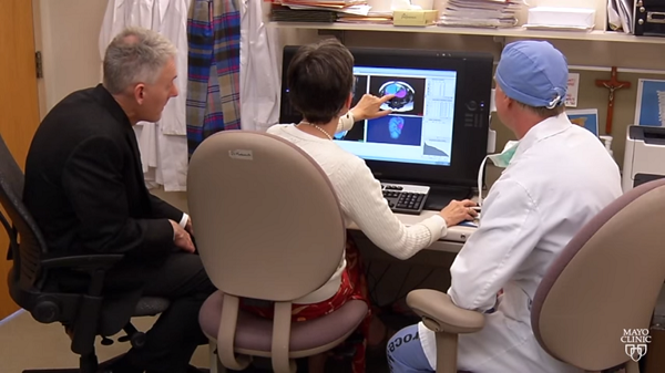
The day of an ultrasound can be one of the most exciting pre-birth connections a parent makes with his/her child. Even though the pictures don’t hold a candle to those taken outside of the womb, there is something amazing about giving form to the life growing in the womb, and they are the first glimpses of the tiny being who will change your world forever. In some cases, however, the information provided by the ultrasound goes beyond the simple introduction of baby to parent and reveals important information about problems that need medical attention. This was the case with an ultrasound performed on expectant mother Caitlin Veitz when she was 20 weeks pregnant.
The ultrasound she received that day would fill the parents with fear and anxiety for their child as it revealed that their baby’s heart was growing outside of the chest wall, a condition that is so rare, it was difficult for the doctors to offer reasonable assurances that they could address the problem. Luckily for her and baby-to-be Kieran, Veitz had access to some of the most highly skilled doctors in the world through the Mayo Clinic. The medical team attending to this special case needed to do everything possible to prepare what Kieran would need, as her very survival was at stake.
An early step in the preparation consisted of the creation of a 3D printed model of Kieran’s anatomy developed from ultrasound data using the Materialise Mimics Innovation Suite. Dr. Jan Matsumoto, radiologist and co-director of the Mayo Clinic’s 3D Anatomic Modeling Lab, then color coded and segmented all of Kieran’s major organs and determined the location of her body in relationship to the placenta, uterus, heart, and liver. It was determined, after working with a life size 3D printed model, that in fact Kieran’s liver and intestines were developing on the outside of her body as well as her heart.
Continue reading in 3dprint.com