
First glimpse of body’s steering wheel joint sparks hope
For the first time, scientists have found a way to reveal the mechanics of the human body’s ‘steering wheel’ – the subtalar joint.
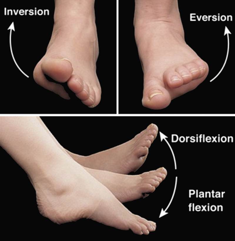
Figure 1.5. Illustration of the inversion/eversion and plantar flexion/dorsiflexion movements. Joint Movements Angular movements increase or decrease the angle between articulating bones. Emile Moreau from Slide 8 Joint Movements Inversion and Eversion of the foot at the ankle, Brian Riley 2016
University of Portsmouth 17 February 2020
The bones of the foot are unique in that they need to be both be extremely flexible allowing the foot to point, twist and flex, but in other positions they need to be absolutely rigid, such as pushing off or jumping so the person doesn’t sprain their ankle.
The key to this ability is the subtalar joint, below the ankle, which until now, doctors couldn’t see rotating while standing.
Ankle sprains are one of the commonest reasons for people to attend Accident and Emergency departments. More often than not, the subtalar joint is also injured but, because the joint is hidden, doctors find it difficult to diagnose sprains, which often leads to long-term ankle instability.
If left untreated, an injury to the subtalar joint can lead to flat feet and even arthritis.
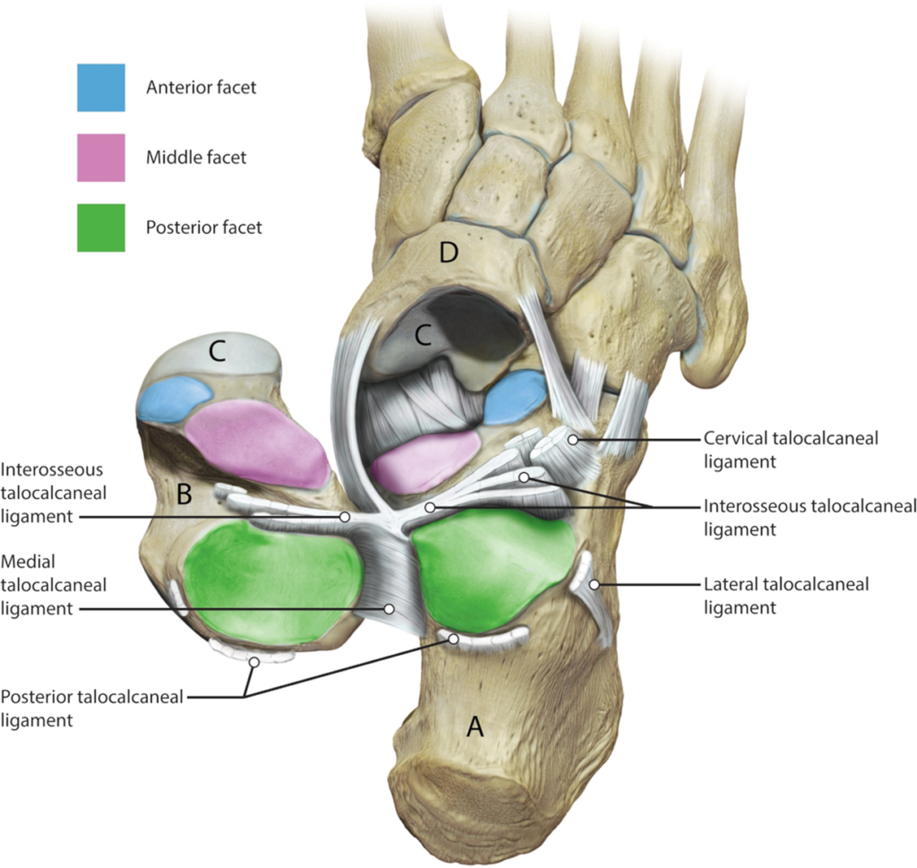
| Figure 1. An illustration of the subtalar joint. The ligaments on the outside of the joint have been divided and the talus (B) has been reflected. The calcaneus (A) is visible from above. The three articular facets of the subtalar joint are illustrated, the posterior facet (green); the middle facet (pink) and the anterior facet (blue). The head of the talus articulates with the navicular bone (D) anteriorly at the talonavicular joint (C). The soft tissue ligamentous restraints are labelled. Image by Catherine Sulzmann, Medical Artist. Fernández, Hoxha, et al. 2020 |
It is hoped that being able to see the joint in action might give doctors the ability to tailor treatments to the many thousands of people with foot joint problems, in the same way it’s possible to tailor treatments of the hip and knee joints.
The study, published in Nature’s Scientific Reports, was led by Dr Gianluca Tozzi, Reader in Bioengineering at the University of Portsmouth, in collaboration with Mr Andrew Goldberg, Consultant Orthopaedic Surgeon at UCL and the Wellington Hospital in London.
Dr Tozzi said: “This is the first time this technique has been used in humans. It is non-invasive and gives clinicians a perfect view of a patient’s subtalar joint motion under full weight-bearing, making it possible for the first time to determine the joint’s centre of rotation which, in turn, opens the possibility of much-improved design of joint replacements.
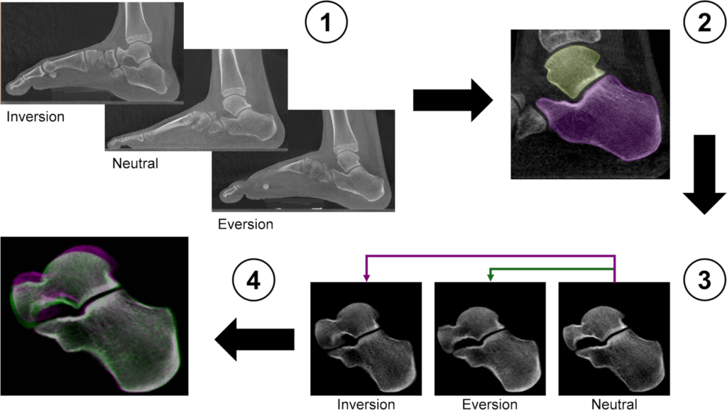
| Figure 9. Workflow of the image post-processing. |
| (1) Weight-bearing clinical CT images of the entire foot in inversion, neutral and eversion positions. |
| (2) Semi-automatic active contour segmentation of the individual in the subtalar joint. |
| (3) The calcaneus in the rotated positions (inversion/eversion) was rigidly register with the corresponding calcaneus in the neutral position. |
| (4) Subtalar joint after rigid registration of the calcaneus. Pink represents inversion and green eversion positions. When pixels of the three configurations match, they displayed the colour grey. After registration, the calcaneus of the three images is perfectly aligned and the relative talus motion can be assessed. Fernández, Hoxha, et al. 2020 |
“Being able to see the subtalar joint in action is made possible by a combination of 3D imaging (computed tomography) and digital volume correlation. The technology has a huge potential to be expanded, allowing doctors to see any strain in the bone, greatly improving clinical diagnosis.
“I’ve always hoped for this. Everyone working in healthcare research hopes their work will be transferable from the lab to real life, making a difference to patients.” – Dr Gianluca Tozzi, Reader in Bioengineering
Mr Goldberg said: “Currently, surgery for arthritis usually involves joining the bones together making them stiff in a procedure known as joint fusion. While this is a successful procedure to treat pain, it does remove a mobile joint which can lead to stiffness and long-term wear of other joints that have to pick up the slack.”
“No one has ever been able to replace this complex joint. This new research helps us to better understand the complex biomechanics of the foot and could pave the way for new treatments that just aren’t currently available.”
 Source University of Portsmouth
| References |
Centre of rotation of the human subtalar joint using weight-bearing clinical computed tomography, Peña Fernández M, Hoxha D, Chan O, Mordecai S, Blunn GW, Tozzi G, Goldberg A. Sci Rep. 2020 Jan 23;10(1):1035. doi: 10.1038/s41598-020-57912-z. Full text
| Further reading |
Study on the three-dimensional orientation of the posterior facet of the subtalar joint using simulated weight-bearing CT, Kleipool RP, Dahmen J, Vuurberg G, Oostra RJ, Blankevoort L, Knupp M, Stufkens SAS. J Orthop Res. 2019 Jan;37(1):197-204. doi: 10.1002/jor.24163. Epub 2018 Nov 29. Full text
Precision of Digital Volume Correlation Approaches for Strain Analysis in Bone Imaged with Micro-Computed Tomography at Different Dimensional Levels, Dall’Ara E, Peña-Fernández M, Palanca M, Giorgi M, Cristofolini L, Tozzi G. Front. Mater. 08 November 2017 https://doi.org/10.3389/fmats.2017.00031 Full text
Micro finite element models of the vertebral body: validation of local displacement predictions, Costa M, Tozzi G, Cristofolini L, Danesi V, Viceconti M, Dall׳Ara E, 11 Jul 2017, PLoS One. 12, 7, e0180151 Full text
Structural and Kinematic Aspects of a New Ankle Rehabilitation Device, Racu (Cazacu) C, Doroftei I. Machines. Technologies. Materials. Vol. 9 (2015), Issue 12, pg(s) 7-10. PDF
In vivo kinematics of the talocrural and subtalar joints with functional ankle instability during weight-bearing ankle internal rotation: a pilot study, Kobayashi T, No Y, Yoneta K, Sadakiyo M, Gamada K. Foot Ankle Spec. 2013 Jun;6(3):178-84. doi: 10.1177/1938640013477452. Epub 2013 Feb 25.
Determination of consistent patterns of range of motion in the ankle joint with a computed tomography stress-test, Tuijthof GJ, Zengerink M, Beimers L, Jonges R, Maas M, van Dijk CN, Blankevoort L. Clin Biomech (Bristol, Avon). 2009 Jul;24(6):517-23. doi: 10.1016/j.clinbiomech.2009.03.004. Epub 2009 Apr 7.
In-vivo range of motion of the subtalar joint using computed tomography, Beimers L, Tuijthof GJ, Blankevoort L, Jonges R, Maas M, van Dijk CN. J Biomech. 2008;41(7):1390-7. doi: 10.1016/j.jbiomech.2008.02.020. Epub 2008 Apr 10.
| Subtalor Joint. Erik Dalton. Vimeo April 14, 2019 |
Also see
Study Reveals The Mechanics of The Body’s ‘Steering Wheel’ For The First Time Science Alert
Subtalar Joint… the body’s steering wheel Erik Dalton Blog
Weight Bearing CT Helps Visualize OCD Lesions CurveBeam