Sever disease: Intervene early to relieve symptoms
Once pain and inflammation have been addressed, clinicians can implement interventions—including orthotic devices, stretching, and strengthening—to address the biomechanical factors that are believed to contribute to heel pain and other symptoms in this population.

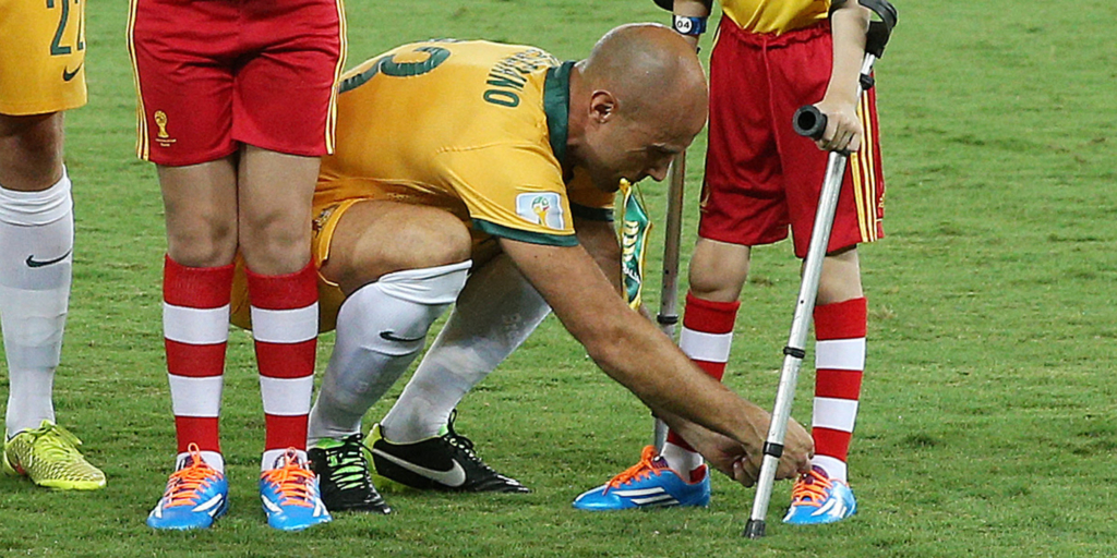
Mark Bresciano of Australia helps tie the shoe laces of a boy ahead of the 2014 FIFA World Cup Brazil Group B match between Chile and Australia at Arena Pantanal on June 13, 2014 in Cuiaba, Brazil. Photo by George Salpigtidis/Newspix/Getty Images.
By Erin Boutwell, Lower Extremity Review May 2015
Sever disease, also known as calcaneal apophysitis, is an overuse injury commonly diagnosed in children, particularly those active in sports such as running and soccer. The primary complaint of Sever disease is heel pain associated with repetitive microtrauma of the calcaneal apophysis. The age range of the children affected is typically 7 to 15 years, corresponding to the time period that starts when the calcaneal growth center appears and ends when it fuses.
Sever disease is considered self-limiting, meaning the disease will resolve itself over time (through fusion of the growth center), but that can take years. Because most patients respond to treatment within three to six weeks and are able to return fully to their normal activities, experts said early intervention is warranted.
“If you have a child who’s in pain for two years of a twelve-year period, that’s a substantial amount of time, so you do want to jump on it and treat it,” said Alicia James, BPod, MHealth Sci, director of the Kingston Foot Clinic in Cheltenham, Australia, who is currently studying interventions in children with Sever disease as a PhD candidate at Monash University in Melbourne, Australia.
Approximately 2% to 16% of the musculoskeletal complaints reported in children are attributed to Sever disease, but these data come primarily from sports medicine clinics and therefore are not necessarily representative of the incidence within the general population.
| Possible mechanisms |
It is widely accepted that Sever disease is the result of microtrauma sustained during repetitive loading. Beyond that, there is no clinical consensus on what causes this microtrauma.
There are two main schools of thought on the biomechanics behind Sever disease. One theory is that the calcaneal apophysis is placed under traction during this period of rapid growth as the Achilles tendon creates a shear force upward on the heel; the plantar fascia may contribute to this traction by pulling in the opposite direction. A second potential explanation for the symptoms of Sever disease is damage caused by the repetitive impact forces during heel strike, particularly during high-impact activities.
Not all Sever patients present with symptoms beyond heel pain. However, some children with Sever disease also present with tight heel cords, limited ankle dorsiflexion, or an underlying biomechanical malalignment.
A prospective study in which investigators compared a group of Sever patients to a control group demonstrated little evidence of these factors, however. The one exception was a significant difference in forefoot position between groups, providing evidence in support of biomechanical malalignment within the Sever disease population.
“Excessive pronation unlocks the foot, making it a mobile adaptor rather than a rigid lever,” said study author Rolf Scharfbillig, PhD, a podiatrist and lecturer at the University of South Australia in Adelaide. “The calf muscles have to work harder to achieve heel lift as [a mobile adaptor] is less mechanically efficient.”
| Erin Boutwell is a freelance writer based in Chicago. |
Continue reading in Lower Extremity Review
Abstract Sever’s disease: what does the literature really tell us? Scharfbillig RW, Jones S, Scutter SD. J Am Podiatr Med Assoc. 2008 May-Jun;98(3):212-23.
Also see
Taller, heavier children have heightened Sever disease risk Lower Extremity Review
