Eagle brace helps slow scoliosis in young – in Calgary
As any parent at bedtime knows, getting kids to do something they don’t want to is one of life’s biggest challenges. In the late 1990s, physicians at the Alberta Children’s Hospital were looking for inspiration on how to coax young patients with scoliosis to wear the uncomfortable braces that can help slow the progression of the disease affecting about two percent of the population.

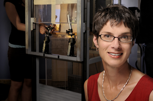
| Dr. Janet Ronsky’s Eagle Brace is helping slow the progression of scoliosis in young patients. The scoliosis brace is named in recognition of the Fraternal Order of Eagles’ generosity. |
What they wanted was a formula to predict the speed and prognosis of scoliosis, a degenerative curvature of the spine, with the ultimate goal of a spinal brace design which could better mold to individual bodies.
They turned to Dr. Janet Ronsky, founding director of the Centre for Bioengineering Research and Education at the University of Calgary and Canada Research Chair in Biomedical Engineering. Her research focuses on joints but spans the fields of engineering, medicine and kinesiology. “When [a brace is] not worn, it can’t correct anything,” she says. So, she and her team created the Eagle Brace.
Scoliosis is more prevalent in girls and often becomes apparent between ages nine and 17. The curvature, which may twist to a helix shape, can dramatically worsen during the growth spurts of adolescence. If left unchecked, the curve can progress to where surgery is required to insert rods and screws—a process which can increase the pain of what is already an excruciating ailment.
| Comfort a factor |
A common treatment is bracing, which holds the torso in place to stop the curve’s growth. But the corset-like device is hot, sweaty and uncomfortable, especially for boys and girls with slim hips. Too often, the child doesn’t want to wear the brace for the upwards of 18 hours a day doctors believe it is necessary to make a difference.

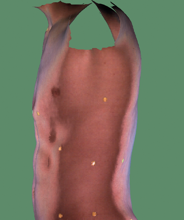
3D scan of torso – The Calgary protocol for the treatment of pectus carinatum. Schulich School of Engineering, University of Calgary.
To address this, U of C engineers worked with researchers at Montreal’s École Polytechnique, using special X-rays to create a three-dimensional image which included the spine’s deformity. A mathematical computation was then used to generate a 3D image of “beads” outlining the youth’s projected form six months down the road, similar to the technique seen on forensic reconstruction TV shows such as Bones.
It worked. Models designed by graduate student Hongfa Wu correctly predicted the curvature advancement within five per cent. This information is then used to design a brace specially built for each patient. The new process also means “we can reduce the number of X-rays we need for monitoring the progression of scoliosis,” says Ronsky.
| This work is part of the University of Calgary’s commitment to expanding its internationally recognized research in the rapidly growing field of biomedical engineering. More than 100 researchers from the Schulich School of Engineering and the faculties of science, medicine, veterinary medicine and kinesiology are involved in inventing, developing and commercializing technologies in the health care sector that will help prevent, diagnose and treat illnesses. The U of C is also creating a new nationally focused innovative centre for biomedical engineering as a catalyst for Alberta’s expanding biomedical economy. |
 Source University of Calgary
The Fraternal Order of Eagles provided the seed money to develop the Eagle scoliosis brace which is used to control the curve of the spine during the growing years. The University of Calgary received a significant donation to Scoliosis research in 2008.
“When we came on board with the project, the young children that suffer from scoliosis had to go in a brace off the shelf which became very, very uncomfortable for them,” said John Potter of the Fraternal order of Eagles.
“I think that a more comfortable brace helping them through this difficult time, or reduction of the maximum dose of radiation to their bodies over the years while we’ve been trying to manage their Scoliosis, I think is a huge step forward,” said Pediatric Orthopedic Surgeon Dr. James Harder.
![]() Source CTV New Calgary, September 7, 2008
Source CTV New Calgary, September 7, 2008
Aeries in Action bulletin, Fraternal Order of Eagles February 2001
| References |
Reconstruction of laser-scanned 3D torso topography and stereoradiographical spine and rib-cage geometry in scoliosis, Poncet P, Delorme S, Ronsky JL, Dansereau J, Clynch G, Harder J, Dewar RD, Labelle H, Gu PH, Zernicke RF. Comput Methods Biomech Biomed Engin. 2000;4(1):59-75.
