
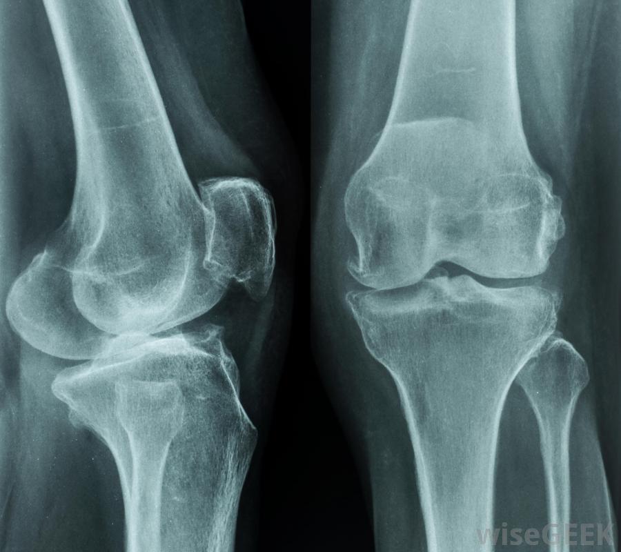
Got knee pain? The best tool for evaluating knee pain among middle-aged patients is an x-ray
Knee pain is common among Americans age 40 and up. Nearly 1 in 17 people visit doctors’ offices each year for knee pain or injuries from osteoarthritis—a progressive “wear and tear” disease of the joints. Those odds increase as the U.S. population continues to age and becomes even more overweight.

The secret to aging: Use it or lose it. The Masters Athlete
Journal of the American Academy of Orthopaedic Surgeons, AAOS September 8, 2016
While a magnetic resonance imaging (MRI) is one tool that can help doctors diagnose torn knee ligaments and cartilage and other problems, plain X-rays are the best first line screening tools for knee pain.
According to a study in the September issue of the Journal of American Academy of Orthopaedic Surgeons, a simple X-ray is frequently the best diagnostic tool, reducing both time and cost.
Whether a patient will need surgery for knee problems depends on how much arthritis he or she has. “If an X-ray shows that a person has significant arthritis, the MRI findings—like a meniscus tear—are less important because the amount of arthritis often dictates the treatment. Therefore, patients should always get a standing X-ray before getting an MRI to screen for knee pain in patients older than 40,” says Muyibat Adelani MD, an orthopaedic surgeon with Washington University’s Department of Orthopedics and lead author of this study.
The study looked at 100 MRIs of knees from patients age 40 and up and found that:
- The most common diagnoses are osteoarthritis (39 percent), and meniscal tears (29 percent)—the tearing of the wedge-shaped pieces of cartilage in the knee joint;
- Nearly 1 of 4 MRIs was taken prior to the patient’s first having obtained a weight-bearing X-ray; and,
- Only half of those MRIs obtained prior to meeting with an orthopaedic surgeon actually contributed to a patient’s diagnosis and treatment for osteoarthritis.
“Patients should always get weight-bearing X-rays before getting an MRI because MRIs are not always needed to diagnose knee problems,” says Dr. Adelani. In cases where arthritis is suspected, weight-bearing X-rays often are more than enough for orthopaedists to complete the diagnosis and treatment plan. An appropriately timed consultation with an orthopaedic surgeon can be more cost effective than first obtaining MRI scans.
Source Journal of the American Academy of Orthopaedic Surgeons, AAOS
What Is Genu Recurvatum? Wise Geek
| Weight-bearing radiographs linked to better clinical decision-making than MRIs for patients with knee pain |
Use of weight-bearing radiographs contributed more to clinical decision-making for patients with knee pain than did the use of pre-referral MRIs, according to results.
by Casey Tingle, Healio September 9, 2016
Researchers documented the presence of a pre-referral MRI or plain radiographic status, the results of weight-bearing radiographs, treatment recommendations and the impact of any pre-referral imaging in 599 patients aged 40 years and older with knee pain.
Overall, results showed 22% of patients underwent MRI before orthopedic evaluation. The most common clinical diagnoses were meniscal tear (38%) and osteoarthritis (37%). Researchers found 58% of patients with pre-referral MRIs had plain radiographic studies before MRI, of which 13% were weight-bearing radiographs. According to results, 45% of patients with pre-referral MRIs had joint space loss and 17% had greater than 50% joint space narrowing.
However, when it came to formulating initial treatment recommendations, researchers noted 63% of the pre-referral MRIs were not useful. Similarly, results showed 5% of the MRIs in patients with pre-referral MRIs and weight-bearing radiographs demonstrating greater than 50% joint space loss contributed to treatment recommendations.
“If an X-ray shows that a person has significant arthritis, the MRI findings — like a meniscus tear — are less important because the amount of arthritis often dictates the treatment,” Muyibat Adelani MD, an orthopedic surgeon in the Department of Orthopedics at Washington University, said in a press release from the American Academy of Orthopaedic Surgeons. “Therefore, patients should always get a standing X-ray before getting an MRI to screen for knee pain in patients older than 40 [years].”
Source Healio
| References |
The Use of MRI in Evaluating Knee Pain in Patients Aged 40 Years and Older, Adelani MA, Mall NA, Brophy RH, Halstead ME, Smith MV, Wright RW. J Am Acad Orthop Surg. 2016 Sep;24(9):653-9. doi: 10.5435/JAAOS-D-15-00681.
| Further reading |
Non-invasive imaging of cartilage in early osteoarthritis, Palmer AJ, Brown CP, McNally EG, Price AJ, Tracey I, Jezzard P, Carr AJ, Glyn-Jones S. Bone Joint J. 2013 Jun;95-B(6):738-46. doi: 10.1302/0301-620X.95B6.31414.
 Sports and joint replacement: Patient perspective should be our priority
 Current thinking in surgical approaches to meniscus repair
 Effects of high heel wear and increased weight on the knee during walking
 Web-based exercise program improves knee arthritis therapy
 Don't screen healthy Canadian kids for developmental delay
 Greater reductions in knee OA pain seen with supportive rather than flexib...