
Not all meniscal pathology is relevant in patients with knee OA
Some—but not all—characteristics of meniscal damage on magnetic resonance imaging (MRI) scans are associated with knee osteoarthritis (OA) severity and two-year progression, according to research from Tufts Medical Center in Boston, MA.

Meniscal pathology in the development and progression of knee osteoarthritis. Aspetar Sports Medicine Journal
By Jordana Bieze Foster, Lower Extremity Review September 2016
As an ancillary project to the Osteoarthritis Initiative, investigators analyzed MRI scans of 465 patients with knee OA (71% had a Kellgren-Lawrence radiographic severity grade of 2 or higher) at two visits, two years apart. Findings were adjusted for each patient’s age, sex, and body mass index.
Meniscal maceration was significantly associated with baseline knee pain, prevalence of end-stage knee OA, and bone marrow lesion (BML) volume, along with the change in BML volume at two years.
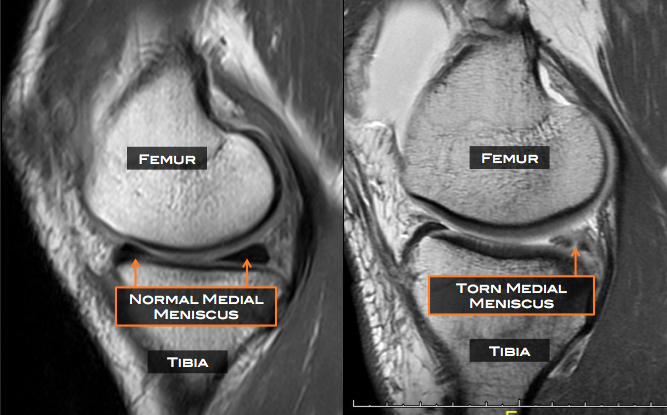
A normal medial meniscus (left) and torn medial meniscus (right) are shown in two MRI examples above. The normal meniscus on these sagittal images (as if viewing from the side) appears as dark black triangles (left). The torn meniscus (right) appears irregular and is traversed by a light line representing the tear. Matt Driscoll MD
Morphological deformity/ extrusion (altered meniscal shape and/or extrusion but no apparent substance loss) was also significantly associated with baseline BML volume and change in BML volume at two years.
Neither intra-meniscal signal nor the presence of meniscal tear were associated with any of the assessed measures of knee OA severity or progression. Osteoarthritis & Cartilage epublished the findings on August 15.
Source Lower Extremity Review
| References |
The relationship between meniscal pathology and osteoarthritis depends on the type of meniscal damage visible on magnetic resonance images: data from the Osteoarthritis Initiative, B Antony, J B Driban, L L Price, G H Lo, R J Ward, M Nevitt, J Lynch, C B Eaton, C Ding, T E McAlindon, Osteoarthritis and Cartilage, Volume 25 , Issue 1 , 76-84. DOI: 10.1016/j.joca.2016.08.004
| Further reading |
Incidental meniscal findings on knee MRI in middle-aged and elderly persons, Englund M, Guermazi A, Gale D, Hunter DJ, Aliabadi P, Clancy M, Felson DT. N Engl J Med. 2008 Sep 11;359(11):1108-15. doi: 10.1056/NEJMoa0800777.
Meniscus pathology, osteoarthritis and the treatment controversy, Martin Englund, Frank W. Roemer, Daichi Hayashi, Michel D. Crema & Ali Guermazi. Nature Reviews Rheumatology 8, 412-419 (July 2012) doi:10.1038/nrrheum.2012.69
Risk factors for medial meniscal pathology on knee MRI in older U.S. adults: A multicenter prospective cohort study, Martin Englund MD PhD, David T. Felson MD MPH, Ali Guermazi MD, Frank W. Roemer MD, Ke Wang MSc, Michel D. Crema MD, John A. Lynch PhD, Leena Sharma MD, Neil A Segal MD MS, Cora E. Lewis MD, and Michael C. Nevitt PhD. Ann Rheum Dis. 2011 October; 70(10): 1733–1739. doi: 10.1136/ard.2011.150052